Human Reproduction vol. 13 no. 3 pp. 509-523, 1998
Sherman J. Silber and Larry Johnson
Tiny numbers of spermatozoa can be extracted from an extensive testis biopsy and be used successfully for intracytoplasmic sperm injection (ICSI) in 60% of cases of nonobstructive azoospermia caused by testicular failure (e.g. maturation arrest, Sertoli cell only, cryptorchid atrophy, postchemotherapy, or even (Klinefelter’s syndrome). However, no sperm are recoverable in 40% of cases even after a very extensive testicular sperm extraction (TESE)-ICSI attempt. Round cells are abundant in morselated testicular tissue of almost all azoospermic men, but difficulties arise in distinguishing under Hoffman or Nomarski optics whether they are haploid round spermatids, diploid spermatocytes or spermatogonia, or even somatic cells like Sertoli cell nuclei or Leydig cells. This paper attempts to clarify such confusion by reviewing data on 143 consecutive testis biopsies of men with non-obstructive azoospermia due to germinal failure, and 62 controls with obstructive azoospermia and normal spermatogenesis. In no cases were round spermatids found in the absence of elongated spermatozoa, and maturation arrest was found always to be a failure of progression beyond meiosis (not at maturation from round spermatid to mature elongated spermatid). Errors arising after injecting somatic or other round cells could result in an appearance resembling fertilization and cleavage, and explain reports of finding ’round spermatids’ in azoospermic men where no ‘spermatozoa’ were retrievable. The use of TESE-ICSI to achieve pregnancies in azoospermic men with deficient spermatogenesis is more concerned with finding tiny foci of spermatozoa rather than searching for ’round spermatids’, which are recoverable only if elongated forms are also available.
Introduction
The discovery that azoospermic men with germinal failure often have minute foci of spermatogenesis, was observed in the early studies of quantitative analysis of spermatogenesis (Steinberger and Tjioe, 1968; Zuckerman et al., 1978; Silber and Rodriguez-Rigau, 1981). However, the importance of this finding for helping azoospermic men with testicular failure have their own genetic child, was not readily apparent until the era of intracytoplasmic sperm injection (ICSI) (Palermo et al., 1992; Van Steirteghem et al., 1993). In 60% of cases of azoospermia caused by testicular failure e.g. maturation arrest, Sertoli cell only, cryptorchid testicular atrophy, post-chemotherapy azoospermia, or even Klinefelter’s syndrome), a tiny number of spermatozoa can often be extracted from an extensive testicle biopsy, and these few retrieved spermatozoa, using ICSI, can result in a normal pregnancy (Devroey et al., 1995; Silber et al., 1995a,b,c, 1996). We termed this procedure TESE (testicular sperm extraction).
However, 40% of azoospermic men with germinal failure have no sperm recoverable during an extensive TESE-ICSI procedure. Recently the possibility has been investigated of using ’round spermatids’, or ’round cells’, derived from testicular tissue (or even from the ejaculate), that are presumably early spermatids, to inject for ICSI for such cases when no elongated spermatozoa are recoverable. Many infertility clinics have attempted ICSI with ROSNI (round spermatid nucleus injection) or ROSI (round spermatid injection). The concept behind this is to provide an option for those patients in whom mature spermatozoa cannot be identified in the TESE-ICSI procedure. Unfortunately there has been a great deel of ignorance and frank deception unfurled on an innocent public regarding the treatment of such couples.

The intention of this scientific paper is to present the outcome of an extensive series of testicle biopsies in all varieties of azoospermic men, to review our previously published histological findings in azoospermic men suffering from ‘maturation arrest’, and to give a view of our attempts to understand the ROSI procedure. It is much easier to discern the various stages and progression of spermatogenesis and of ‘spermiogenesis’ in well-prepared, stained histological slices than the unstained cytological specimens found at TESE-ICSI. Photographs are included to help in vitro (IVF) laboratory personnel to identify the many various types of ’round cells’ seen in a dissected testicular specimen, elying to some extent not only on present efforts, but also on well established previously published reports.
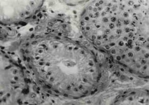
The tubule in the center left, as well as upper and lower far left, shows only Sertoli cells which are often confused by inexperienced enthusiasts with “round” spermatids, but in truth represent only the nourishment cells of the testis. The tubule on the upper right has normal sperm production with all stages of spermatogenesis present. The secret to obtaining specimens from men who appear to be making no sperm is not to inject round cells, but rather through microsurgery to locate the true sperm, or the mature, oval, condensed spermatids.
Histological examinations of testicle biopsy slides in patients with non-obstructive and obstructive azoospermia
Our library of testicular histology for non-obstructive azoospermia due to germinal failure totalled 143 patients. Sixty-six of these 143 men had a diagnosis of ‘Sertoli cell only’ with no other known cause for infertility. Fifty-nine of these patients had a diagnosis of either pure ‘maturation arrest’ or a combination of ‘maturation arrest’ and ‘Sertoli cell only’ in different areas of the same slide. Eighteen patients had other causes for non-obstructive ‘ azoospermia, such as mumps orchitis, sex chromosomal anomaly, cryptorchidism, or previous chemotherapy.
For controls with ‘normal spermatogenesis’ we used 62 men with obstructive azoospermia, caused either by congenital absence of the vas or vasoepididymostomy failure, who over the course of the last 3 years, have undergone TESE-ICSI procedures (because of either an absence of epididymis or a failure to find motile spermatozoa in the epididymis). Thus, we were able to review a library consisting of 205 testes biopsies, 143 having non-obstructive azoospermia due to testicular failure, and 62 with obstruction and normal spermatogenesis. These slides were all reviewed in a quantitative fashion as has already been described (Steinberger and Tjioe, t 1968; Zuckerman et al., 1978; Silber and Rodriguez-Rigau, l98l; Johnson et al., 1992). We found no cases of classic ‘hypospermatogenesis’ (as we define it) in azoospermic patients. Classic hypospermatogenesis implies a diffuse reduction in quantitative spermatogenesis throughout the testis and it is generally associated with oligozoospermia, not azoospermia.
Figures 1A,B and 2A-C illustrate the findings of spermatogenesis genesis in all categories of non-obstructive azoospermia studied. In the case of Sertoli cell only, there is, of course, an absence of germ cells. If one looks at an entire slide of more than 20 tubules with ‘Sertoli cell only.’ in many cases there will be an occasional tubule with normal spermatogenesis (Silber, 1995b; Silber et al., 1996). The minimum number of tubules counted on both sides was 40 per patient, and usually >100 tubules were counted.
The Sertoli cell is a large, formless amoeba-like cell in which the germ cells would normally be nourished. The nuclei of the Sertoli cells are located along the basement membrane circumferentially at the base of the seminiferous tubule, and each contains a very prominent nucleolus. The Sertoli cell nucleus is a dominant presence in the histology of Sertoli cell only. It is important to keep this picture in mind when searching for ’round spermatids’ in men with no spermatozoa found at TESE-ICSI.
Figure 3 shows representative histology from a patient with ,maturation arrest’. In all cases, the arrested development was found to occur in meiosis, either at zygotene or pacbytene. No round spermatids were found except in those cases (partial) where elongated spermatids and mature spermatozoa also occurred. Thus, in none of the 125 cases of idiopathic nonobstructive azoospermia was there any evidence of ‘spermiogenic’ arrest, i.e. arrest in the development of mature elongated spermatids from round spermatids, The germinal defect in non-obstructive azoospermia, as already reported, was either an absence of germ cells (Sertoli cell only), or a failure of germ cells to progress beyond meiosis (maturation arrest) (Silber et al., 1996).
In the other miscellaneous causes of non-obstructive azoospermia, whether from chemotherapy, cryptorchidism, or mumps, we found varying degrees of fibrosis and tubular atrophy that were not seen in the idiopathic examples previously discussed. However, once again the defect in spermatogenesis in all these cases involved either an absence of germ cells, or a failure of the germ cells to progress beyond meiosis. Thus, in all 143 cases of non-obstructive azoospermia caused by testicular failure, spermiogenic arrest, i.e. failure of round spermatids to develop into mature spermatozoa, was never found. Such a condition must be fairly uncommon.
Figure 4A-C shows examples of the histology of 62 patients with obstructive azoospermia undergoing TESE-ICSI, who presumably should have had normal spermatogenesis. All of these cases with a clinical diagnosis of obstruction had full progression of spermatogenesis, both premeiotic and postmeiotic. It is readily apparent that zygotene and pachytene spermatocytes are somewhat bigger than round spermatids, but Sertoll cell nuclei, leptotene spermatocytes and the briefly present secondary spermatocytes are all of similar size to round spermatids.
It would appear that in some tubules which exhibit normal spermatogenesis, there is a predominance of round spermatids, but a review of many seminiferous tubules in these cases still revealed a normal progression of round to mature spermatids (Johnson et al., 1992). It has long been known that in humans there is no orderly wave of progressive spermatogenic stages down the seminiferous tubules, as in most animals (Clermont, 1972). Therefore, the appearance of just a few tubules is not representative of the rest of the testicle, but of twenty or more tubules is. In these 205 cases of testicle biopsies in azoospermic men, we were not able to find any tubules in which round spermatids were observed in the absence of mature spermatozoa.
An atlas of male germ cells: to be used for identifying round spermatids during a TESE-ICSI procedure
When one performs a TESE-ICSI procedure in patients with testicular failure, as well as in patients with normal spermatogenesis, there is always an abundance of ’round cells’. It is very difficult with Hoffman optics to differentiate with certainty a round spermatid from a Sertoli cell nucleus with its prominent nucleoIus, or even from a spermatocyte. Even when there are truly no spermatozoa at all, there will always be many ’round cells’ seen with either Sertoli cell only or with maturation arrest, but these are not round spermatids (Johnson et al., 1981, 1992; Johnson, 1986; Silber et al., 1996). Figure 5A,B was taken from our collection of TESE cases.
With Hoffman and Nomarski optics normally used with ICSI, it is very difficult to distinguish round spermatids from Sertoli cell nuclei. The round spermatid should be distinguished by the acrosomal vesicle located on the periphery, and this does not show up well on Hoffman or Nomarski optics. In Figure 6 the round spermatid can be distinguished by the ‘glow’ of the early acrosomal cap (Holstein and Roosen-Runge, 1981). This can only be reliably and simply visualised with phase contrast.
Clinical experience with ROSI and ROSNI
The first reports of success (Tesarik et al., 1995, 1996; Tesarik and Mendoza, 1996) described seven cases of azoospermic men, which, despite the absence of mature spermatozoa, had round spermatids in the ejaculate, and these round spermatids were injected (instead of mature spermatozoa), resulting in two out of seven successful pregnancies with viable births. This would be an incredible increase in efficiency compared to the 1% live birth rate that Ogura and Yanagimachi obtained in mice injected with round spermatids.
Perhaps more importantly, the finding of round spermatids in the absence of mature spermatozoa in an azoospermic man appears to contradict our observation that in humans, round spermatids are not found in the absence of mature spermatids. Nonetheless, a few other centres have made similar claims (Sofikitis et al., 1996. Antinori et al., 1997a, b; Fishel et al., 1997).
A further report of the use of spermatid injection came from Fishel et al. (1995). Their successful pregnancy resulted from injection of a spermatid retrieved from the testicle, rather than the ejaculate, but these were not to round spermatids. The author suggests that spermatozoa were not available and, therefore, they had to resort to choosing earlier spermatids. However, in this case report, some of the ejaculates of the patient actually had a few spermatozoa (crypt-azoospermia), and other ejaculates were azoospermic. An ill-conceived attempt apparently was made to retrieve spermatozoa from the epididymis (rarely successful in non-obstructive azoospemia), but then finally a testicular biopsy was performed and an attempt at TESE was made. Apparently, pine spermatozoa were actually recovered, but the morphology was deemed by the authors to be abnormal and, therefore, instead they chose to inject what they called ‘elongated spermatids,’ which looked ‘healthier than the few spermatozoa obtained.’ It is obvious that these ‘spermatids’ were so mature that they were in truth normal mature spermatozoa.
Although Fishel et al. (1995) discuss the ’round spermatid’ injection, clearly what they were reporting is no different from the routine sperm injections that have been reported already for non-obstructive azoospermia (Devroey et al., 1995; Silber et al., 1995a; 1996). Similar reports of ‘late spermatid injection’ have been made by Vanderzwalmen et al. (1995) and Araki et al. (1997). However, most of these successful cases are just sporadic reports of what is no different than simply TESE-ICSI with ‘elongated spermatids’, i.e. testicular sperm extraction, finding occasional spermatozoa present in 60% of testicle specimens from men with azoospermia caused by germinal failure.
Nonetheless, there is still a great deal of interest in attempting round cell injection in cases of azoospermia where no spermatozoa or elongated spermatids are found during the TESE procedure. However, it is very difficult to decide what really constitutes a round spermatid, a secondary spermatocyte, a primary spermatocyte, and even spermatogonia, Leydig cells and Sertoli cell nuclei in the usual ICSI setting. This has been the bais of many deceptive practices on the part of some clinics. Non-specific egg activation could serve as asource of confusion to some enthusiasts for ROSI and ROSNI.
It would appear that where true round spermatids are found, mature spermatids should also be retrievable, and would certainly be preferable for injection.
Conclusion
One of the problems for lVF clinics using the TESE-ICSI procedure is that the embryologist and clinician may possibly have little input from either a urologist or an endocrinologist who is experienced with spermatogenesis and testicular histology. Our discovery that small numbers of spermatozoa sufficient for ICSI can be found in the testes of azoospermic men, does not mean that the testicle is a matzoh ball full of spermatozoa. and round cells just waiting for injection.
We conclude that the ability to use TESE-ICSI to achieve pregnancies and babies in azoospermic men with deficient spermatogenesis is related to the ability to find tiny foci of spermatozoa in a testicle that otherwise is grossly deficient in spermatogenesis (such that not enough spermatozoa are being produced to reach the ejaculate), and not upon the ability to find less ‘mature forms such as ’round spermatids’ in these patients.
References
- Antinori. S., Versaci. C., Dani. G. et at. (1997) Fertilization with human testicular spermatids: four successful pregnancies. Hum. Reprod., 12. 286-291.
- Araki, S., Motoyama, M., Yoshida, A. et at. (1997) Intracytoplasmic injection with later spermatids: a successful procedure in achieving childbirth for couples in which the male partner suffers from azoospermia due to deficient spermatogenesis. Fertil. Steril., 67, 559-561.
- Clermont, Y. (1972), Kinetics of spermatogenesis in mammals; seminiferous epithelium cycles and spermatogonial renewal. Physiol. Rev., 52, 198-236.
- Devroey, P., Liu, J.. Nagy, Z. et al. (1995) Pregnancies after testicular sperm extraction (TESE) and intracytoplasmic, sperm injection (ICSI) in nonabstractive azoospermia. Hum. Reprod., 10, 1457-1460.
- Edwards, R.G., Tarin, J.J., Dean. N. et al. (1994) Are spermatid injections into human oocytes now mandatory? Hum. Reprod., 9, 2217-2219.
- Fishel, S., Green, S., Bishop, M. et al. (1995) Pregnancy after intracytoplasmic injection of spermatid. Lancet, 345, 641-642.
- Holstein. A.F. and Roosen-Runge. E.C. (eds) (1991) Atlas of Human Spermatogenesis. Grosse Verlag, Berlin.
- Johnson, L. (1986) Review article. Spermatogenesis and aging in the human. J. Androl., 7. 331-354.
- Johnson. L.. Pert ” . C.S. and Neaves, W.B. (1981) A new approach to quantification of spermatogenesis and its application to germinal cell attrition during human spermiogenesis, Biol. Reprod., 25, 217-226.
- Johnson. L., Chaturedi. P.K. and Williams. J.D. (1992) Missing generations of spermatocytes and spermatids in seminiferous epithelium contribute to low efficiency of spermatogenesis in human. Biol. Reprod., 47, 1091-1099.
- Kimura. Y. and Yanagimachi. R. (1995) Mouse oocytes injected with testicular spermatozoa or round spermatids can develop into normal offspring, Development, 121. 2397-2405.
- Mendoza, C. and Tesarik, J. (1996) The occurrence and identification of round spermatids in the ejaculate of men with non-obstructive azoospermia. Fertil. Steril., 66, 826-829.
- Mendoza, C., Benkhalifa. M., Coben-Bacrie. P. et al. (1996) Combined use of proacrosin immunocytochemistry and autosomal DNA in situ hybridisation for evaluation of human ejaculated germ cells. Zygote, 4, 279-283.
- Ogura, A. and Yanagimachi, R. (1993) Round spermatid nuclei injected into hamster oocytes form pronuclei and participate in syngomy. Biol. Reprod., 48, 219-225.
- Ogura. A., Yanagimachi, R. and Usui, N. (1993) Behavior of hamster and mouse round spermatid nuclei incorporated into mature oocytes by electrofusion. Zygote, 1, 1-8.
- Ogura, A., Matsuda. J. and Yanagimachi. R. (1994) Birth of normal young after electrofusion of mouse oocytes with round spermatids. Proc.\Natl. Acad. Set. USA, 91. 7460-7562.
- Palermo, G., Joris, H., Devroey, P. and Van Steirteghem, A. (1992) Pregnancies after intracytoplasmic injection of single spermatozoan into an oocyte. Lancet, 340, 17-18.
- Silber, S.J. and Rodriguez-Rigau, L. (198 1) Quantitative analysis of testicle biopsy: determination of partial obstruction and prediction of sperm count after surgery for obstruction. Fertil. Steril., 36, 480-485.
- Silber. S.J., Van Steirteghem, AC and Devroey. P. (1995a) Sertoli cell only revisited. Hum. Reprod., 10, 1031-1032.
- Silber, S.J., Van Steirteghem, A.C., Liu, 1. et al. (1995b) High fertilization and pregnancy rate after intracytoplasmic sperm injection with sperm obtained from testicle biopsy. Hum. Reprod., 10, 148-152.
- Silber, S.J., Nagy, Z., Liu, J. et al. (1995c) The use of epididymal and testicular spermatozoa for intracytoplasmic sperm injection: the genetic implications for male infertility. Hum. Reprod., 10, 2031-2043.
- Silber, ST. Van Steirteghem, A.C., Nagy, Z. et al. (1996) Normal pregnancies resulting from testicular sperm extraction and intracytoplasmic sperm injection for azoospermia due to maturation arrest, Fertil. Steril., 66. 110-117.
- Sofikitis N.V., Miyagawa, I., Agapitos, E. et al. (1994) Reproductive capacity of the nucleus of the male gamete after completion of meiosis. J. Assist. Reprod. Genet., 11, 335-341.
- Sofikitis, N.. Toda. T., Miyagawa, I. et al. (1996) Beneficial effects Of electrical stimulation before round spermatid nuclei injections into rabbit oocytes on fertilization and subsequent embryonic development. Fertil. Steril., 65. 176-185.
- Steinberger, E. and Tjioe, D.Y. (1968) A method for quantitative analysis of human seminiferous epithelium. Fertil. Steril., 19, 960-970.
- Tesarik, J. and Mendoza, C. (1996) Spermatid injection into human oocytes. 1. Laboratory techniques and special features of zygote development. Hum. Reprod., 11, 772-779.
- Tesarik, J., Mendoza, C. and Testart, J. (1995) Viable embryos from injection of round spermatids into oocytes (Letter). N. Engl. J. Med., 333, 525.
- Tesarik, J., Role[, F., Brami, C. et al. (1996) Spermatid injection into human oocytes. 11. Clinical application in the treatment of infertility due to nonabstractive azoospermia. Hum. Reprod., 11. 780-783.
- Vanderzwalmen Is, Lejeune, B., Nijs, M. et al. (1995) Fertilization of an oocyte microinseminated with a spermatid in an in vitro fertilization programme. Hum. Reprod., 10. 502-503.
- Van Steirteghem, A.C., Nagy, Z., Joris, H. el al. (1993) High fertilization and implantation rates after intracytoplasmic sperm injection, Haiti. Reprod., S. 1061-1O66.
- Verheyen, G., Crabbe, E., Joris, H. and Van Steirteghem. A. (1998) Simple and reliable identification of the human round spermatid by invented phasecontrast microscopy; Does the real target group exist? Man. Reprod., 13, in press.
- Zuckerman, Z., Rodriguez-Rigau. L.J., Weiss, D.B. et al. ( 1978) Quantitative analysis of the seminiferous epithelium in human testicle biopsies and the relation of spermatogenesis to sperm density. Fend. Steril., 30, 448-455
